Canine atopic dermatitis: a roadmap to individualized, multimodal treatment

Mary Fuller, DVM
Director of Veterinary Services

In the last decade, new drug therapies have transformed the way veterinarians treat canine atopic dermatitis (AD). Clinicians are now able to bring dogs fast relief from pruritus and inflammation with fewer adverse effects. Still, treatment remains challenging.
In an attempt to provide veterinarians with sound guidance, treatment guidelines for canine atopic dermatitis were developed in 2010 by the International Committee on Allergic Diseases of Animals (ICADA) and have subsequently been updated in 2015.1,2
Disease overview
Atopic dermatitis is a common canine skin disorder estimated to affect about 10 to 15% of dogs.3 Affected canines are genetically predisposed to develop inflammatory and pruritic skin lesions associated with IgE antibodies targeted against environmental allergens.4
Signs typically develop early in life, between 6 months and 3 years of age. Distribution of lesions can vary, but may include the axillae, abdomen, inguinal, perineum, face, concave pinnae, as well as the dorsal and ventral paws.1 Depending on the allergens involved, clinical signs may be seasonal or non-seasonal, and the disease typically requires lifelong management.
Pathogenesis of AD
“The pathogenesis [of canine atopic dermatitis] is very complex and involves multiple organ systems including the skin, the immune system, and the nervous system,” according to Douglas DeBoer, DVM, DACVD, Professor at the School of Veterinary Medicine at the University of Wisconsin-Madison.
While AD was once believed to be a hypersensitivity to inhaled environmental allergens, there is now growing evidence that epidermal barrier defects may play an important role.5
Environmental allergens are thought to penetrate the epidermis, where they are captured by antigen-presenting cells. These, in turn, travel to the dermis and lymph nodes, where they activate T-helper 2 lymphocytes. These cells release cytokines or inflammatory mediators that boost allergen-specific IgE antibodies as well as pruritogenic cytokines such as interleukin-31 (IL-31) that directly stimulate nerves to promote itching.
“All of the pathways that cause inflammation and pruritus are orchestrated through the action of cytokines,” according to Dr. DeBoer in a presentation at the 2020 VMX conference.6
An individualized, multimodal approach
Traditional treatment of AD primarily relied on systemic glucocorticoids. While these medications inhibited the production of many cytokines associated with itching and inflammation, they were non-targeted in that they also inhibited other types of cytokines. This often resulted in adverse effects associated with the adrenal glands, liver, and kidneys.
To help minimize potential adverse effects, clinicians now tend to favor a multimodal approach, using a combination of products to produce synergistic effects.
The AD treatment roadmap
Once a diagnosis of canine AD has been confirmed, ICADA guidelines generally focus on treatment of acute flares or chronic AD and vary by whether signs are localized or generalized. The following is a brief summary of ICADA recommendations.1,2
Identify and avoid flare factors
ACUTE FLARES AND PREVENTION OF RECURRENCE1,2
- Determine if there are flare factors at play, including environmental allergens (especially house dust mites and pollens), food allergens, or fleas, and remove the source, if possible.
- Treat bacterial and yeast infections of the skin and ears with appropriate and responsible antimicrobial therapy. Medicated shampoos [eg, KETOCHLOR® (Chlorhexidine Gluconate, Ketoconazole) Medicated Shampoo] are recommended. If using topical antimicrobials, clinicians should monitor the skin for drying or irritation, which may also cause flares.
CHRONIC AD1,2
- Flea control: Dogs with AD can be predisposed to flea bite hypersensitivity if repeatedly exposed to flea bites.7 In regions where fleas are endemic, dogs should be on year-round flea adulticides and owners should be instructed on environmental measures.
- Frequent shampooing may impact the efficacy of topical flea control products.
- Environmental allergens: Perform allergen- specific intradermal and/or IgE serologic tests to identify possible environmental allergens or for use in allergen-specific immunotherapy.
- House dust mite control: Glycoproteins from Dermatophagoides house dust mites are the most common allergens in dogs globally.8 Reducing these in the dog’s environment may help reduce signs. This may include frequent pet bed washing and environmental vacuuming.
- Food allergy: In dogs with non-seasonal AD, perform 1 or more 8-week dietary restriction-provocation trials to determine if food allergens contribute to clinical signs.
- Bacterial and fungal infections: Staphylococcal and Malassezia skin and ear infections are common in dogs with AD. Some atopic dogs can develop an IgE hypersensitivity to Malassezia9-12 and Staphylococcus.13 Dogs should be treated with topical or systemic antimicrobials as needed.
Improve skin and coat hygiene and care
ACUTE FLARES1,2
- Bathing: A weekly bath, including a 10-minute soak with an emollient shampoo that contains certain lipids, antiseptics, and complex sugars, or raspberry oil, can help remove allergens and reduce itching and lesions. In a randomized controlled trial, 25% of dogs given a weekly bath and 10-minute application with shampoo [ALLERMYL® (Piroctone Olamine) Medicated Shampoo, Virbac] experienced a 50% reduction in pruritus scored within 24 hours.14
CHRONIC AD1,2
• Bathing: A weekly bath with a non-irritating shampoo and lukewarm water is advised.
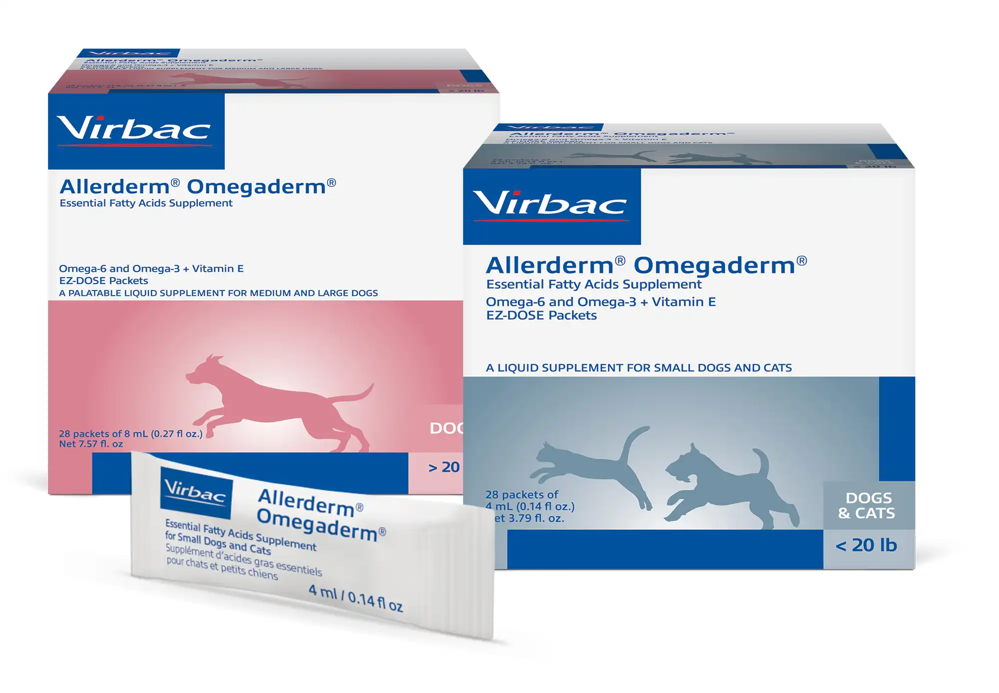
• Oral essential fatty acids (EFAs): Diets or supplements, especially those rich in Omega-6 fatty acids, can help improve skin lipids and coat quality.
Reduce pruritus and inflammation with pharmacological agents
ACUTE FLARES1,2
• Topical glucocorticoid sprays: For control of pruritis associated with AD, 0.015% triamcinolone acetonide (GENESIS® Topical Spray, Virbac) has been shown to be highly effective.15,16
• Oclacitinib: A short course of therapy can help provide quick relief for pruritus and skin lesions, and appears to be safe.
• Lokivetmab: Released after the publication of the 2015 guidelines, this injectable treatment can provide long-lasting relief from clinical signs.
• Oral glucocorticoids: A short course of prednisolone, prednisone, or methylprednisolone can be used for dogs with acute, severe or generalized AD.
• Antihistamines: Oral type 1 antihistamines may provide a small amount of relief to dogs with mild AD, especially if administered before a flare to prevent histamine release.
CHRONIC AD1,2
• Topical glucocorticoid sprays: After daily induction phase, apply intermittently (2 times a week) to prevent recurrence of clinical signs.16
• Oral cyclosporine: With a long history of use, cyclosporine can help induce remission, then the dose or frequency can be tapered for AD maintenance therapy.
• Oral oclacitinib: These pills can be prescribed for acute or chronic control of AD.
• Lokivetmab: A single injection, administered once every 4 to 6 weeks, is also appropriate for acute or chronic management of AD.
• Oral glucocorticoids: Prednisolone, prednisone, or methylprednisolone can help induce remission of clinical signs, and should then be tapered to the lowest effective dose.
Topical and oral steroids
In most cases, broad-spectrum anti-inflammatory action is needed to get an acute flare under control before long-term maintenance therapy can be initiated.16 In dogs, topical steroids are also the cornerstone for inducing remission in acute flares and localized lesions. DeBoer promoted the topical use of “steroids in spray formulation” such as 0.015% triamcinolone acetonide that contain a relatively low concentration of the drug, but offer a low potential for adverse effects in relation to their anti-inflammatory potency.
If clinical signs are severe or more generalized, a short course of oral steroids such as prednisolone, prednisone, or methylprednisolone can be used with topical steroids until control has been achieved.
Cyclosporine
Oral cyclosporine is more targeted than oral glucocorticoids. It inhibits the intracellular enzyme calcineurin, which blocks the production of cytokines, such as interleukin-2 (IL-2). This, in turn, inhibits T-cell activation and proliferation to provide anti-inflammatory and immunomodulatory effects. Modified cyclosporine is available in capsule form. However, 2 randomized, controlled studies showed that a novel liquid formulation of cyclosporine [CYCLAVANCE™ (cyclosporine oral solution) USP MODIFIED, Virbac] was better accepted by dogs than cyclosporine capsules, which may help improve compliance.17,18
Oclacitinib
Oclacitinib has a different mechanism of action than glucocorticoids or cyclosporine. This oral medication selectively inhibits Janus kinase 1 (JAK1) enzymes within cells, blocking JAK1-dependent cytokine activity, including interleukin-31 (IL-31), a cytokine primarily responsible for sending itch signals to the brain. Since oclacitinib typically relieves itch quickly (in a matter of hours), it can be a good choice for acute pruritus. Oclacitinib typically has few adverse effects, but should not be used in dogs less than one year of age. Some recent studies have shown that the use of a topical steroid in conjunction with oclacitinib can reduce the time for remission of signs, and avoid “rebound” flare ups.19
Lokivetmab
Lokivetmab: This injectable monoclonal antibody treatment is even more targeted than oclacitinib, exclusively blocking the action of IL-31. After a subcutaneous injection, lokivetmab typically relieves pruritus in 1 to 3 days and lasts for 4 to 6 weeks. As a biological therapy, lokivetmab isn’t metabolized like a drug, so it’s not likely to cause organ toxicity.
Progressive therapy
“If you have a patient that’s been unresponsive or difficult to treat previously, first verify you have infection and parasite control on board,” advised Dr. DeBoer. He then promoted the concept of progressive therapy, which recognizes that different stages of AD may require different treatments over the course of a dog’s life.
Combining several products can also help manage more of the inflammatory process. For instance, it may be necessary to combine therapies, such as oclacitinib and lokivetmab to provide the dog and owner with immediate itch relief in an acute flare. Other dogs may require a combination of treatments to achieve fast results and to help keep the pet in remission. For example, oclacitinib can be used along with lokivetmab or a topical steroid; or, cyclosporine can be given at the same time a topical steroid is used.
Adapted from an article published by DVM 360
IMPORTANT SAFETY INFORMATION
CYCLAVANCE™ (cyclosporine oral solution USP MODIFIED): For use in dogs only. Wear gloves during and wash hands after administration. Gastrointestinal problems and gingival hyperplasia may occur at the initial recommended dose of CYCLAVANCE oral solution. CYCLAVANCE oral solution should be used with caution: 1) in cases with diabetes mellitus as it may cause elevated levels of serum glucose; 2) in dogs with renal insufficiency since the effect of cyclosporine use on dogs with compromised renal function has not been studied; 3) in simultaneous administration with drugs that suppress the P-450 enzyme system, such as azoles (e.g. ketoconazole), that may lead to increased plasma levels of cyclosporine. Killed vaccines are recommended for dogs receiving CYCLAVANCE oral solution because the impact of cyclosporine on the immune response to modified live vaccines has not been evaluated. For full prescribing information, contact Virbac at 1-800-338-3659 or visit us.virbac.com.
GENESIS® (0.15% triamcinolone acetate) Topical spray: For use on dogs only. Wear gloves when applying the product. The use of this product on dogs less than eight pounds, less than one year of age, breeding, pregnant, or lactating has not been evaluated. Adverse events of polyuria and polyphagia have been reported in <6% of dogs receiving treatment. For full prescribing information, contact Virbac at 1-800-338-3659 or visit us.virbac.com.
REFERENCES
1. Olivry T, DeBoer DJ, Favrot C, et al. Treatment of canine atopic dermatitis: 2010 clinical practice guidelines from the International Task Force on Canine Atopic Dermatitis. Vet Dermatol. 2010;21:233–248.
2. Olivry T, DeBoer DJ, Favrot C, et al. Treatment of canine atopic dermatitis: 2015 updated guidelines from the International Task Force on Canine Atopic Dermatitis. BMC Vet Res. 2015;11:210. doi: 10.1186/ s12917-015-0514-6.
3. Hillier A, Griffin CE. The ACVD task force on canine atopic dermatitis (I): incidence and prevalence. Vet Immunol Immunopathol. 2001;81(3–4):147–151. doi: 10.1016/S0165-2427(01)00296-3.
4. Halliwell R. Revised nomenclature for veterinary allergy. Vet Immunol Immunopathol. 2006;114:2007–2008.
5. Marsella R, Samuelson D. Unraveling the skin barrier: a new paradigm for atopic dermatitis and house dust mites. Vet Dermatol. 2009;20:533–540.
6. DeBoer DJ. New therapies for canine pruritus: How do I choose? VMX2020. Available at: www.vetfolio. com/courses/new-therapies-for-canine-pruritus-how-do-i-choose. Accessed June 3, 2021.
7. Sousa CA, Halliwell REW. The ACVD task force on canine atopic dermatitis (XI): the relationship between arthropod hypersensitivity and atopic dermatitis in the dog. Vet Immunol Immunopathol. 2001;81:233–238.
8. Hill PB, DeBoer DJ. The ACVD task force on canine atopic dermatitis (IV): environmental allergens. Vet Immunol Immunopathol. 2001;81:159–168.
9. Morris DO, Olivier NB, Rosser EJ. Type-1 hypersensitivity reactions to Malassezia pachydermatis extracts in atopic dogs. Amer J Vet Res.1998;59:836–841.
10. Nutall TJ, Halliwell REW. Serum antibodies to Malassezia yeasts in canine atopic dermatitis. Vet Dermatol. 2001;12:327–332.
11. Morris DO, DeBoer DJ. Evaluation of serum obtained from atopic dogs with dermatitis attributable to Malassezia pachydermatis for passive transfer of immediate hypersensitivity to that organism. Am J Vet Res. 2003:64:262–266.
12. Farver K, Morris DO, Shofer F, et al. Humoral measurement of type-1 hypersensitivity reactions to a commercial Malassezia allergen. Vet Dermatol. 2005;16:261–268.
13. Morales CA, Schultz KT, DeBoer DJ. Antistaphylococcal antibodies in dogs with recurrent staphylococcal pyoderma. Vet Immnol Immunopathol. 1994;42:137–147.
14. Löflath A, von Voights-Rhetz A, Jeeger K, et al. The efficacy of a commercial shampoo and whirlpooling in the treatment of canine pruritus—a double-blinded, randomized, placebo-controlled study. Vet Dermatol. 2007;18:427–431.
15. DeBoer DJ, Schafer JH, Salsbury CS, et al. Multiple-center study of reduced-concentration triamcinolone topical solution for the treatment of dogs with known or suspected allergic pruritus. Am J Vet Res. 2002;63:408–413.
16. Lourenco AM, et al. Efficacy of proactive long-term maintenance therapy of canine atopic dermatitis with 0.0584% hydrocortisone aceponate spray: a double-blind placebo controlled pilot study. Vet Dermatol. 2016;27:88–e25. doi: 10.1111/vde.12285.
17. Navarro Crates N, Benizeau E, McGahie D. Voluntary acceptance and consumption of two oral ciclosporin formulations in dogs: two randomized, controlled studies. 2015;68:3. doi: 10.1186/ s13620-015-0031-8.
18. Navarro C, Séguy L, Vila M, Birckel P. Bioequivalence study between two formulations of ciclosporin A (Cyclavance® oral solution and Atopica® soft capsules) following a single oral administration in dogs. BMC Vet Res. 2016;12:54. doi: 1186/s12917-016-0669-9. PMID: 26970736; PMCID: PMC4789266.
19. Takahashi J, Kanda S, Iyori K. Efficacy and safety of a 0.0584% hydrocortisone aceponate spray to reduce flares of canine atopic dermatitis when tapering oclacitinib: a randomized, doble-blinded, placebo-controlled trial. Presented at: European Veterinary Dermatology Congress; 26-28 September 2019; Liverpool, UK.
© 2023 Virbac Corporation. All Rights Reserved. CYCLAVANCE, GENESIS, ALLERMYL, and KETOCHLOR are registered trademarks of the Virbac Group of companies